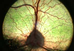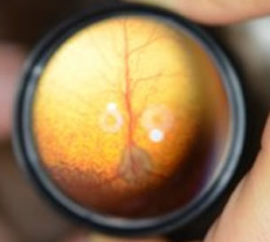

Therapy of inflammatory type posterior segment disease should aim to treat the underlying cause wherever possible (e.g. The retinas are often totally detached and hemorrhage is common.

Cats typically present with a sudden onset of blindness accompanied by dilated non-responsive pupils. Aspirates from the posterior segment should be a last resort.Ī specific retinopathy commonly seen in older cats is hypertensive retinopathy, which may occur secondary to hyperthyroidism, chronic renal failure, and adrenal neoplasia or occur as primary disease. Ultrasonography of the posterior segment is useful where opacities of the anterior segment media or lens obscure a view of the fundus. Workup involves a thorough physical and ocular examination, minimum database, serology and/or PCR for infectious diseases, chest/abdominal radiographs and ultrasound, neurological examination, blood pressure measurement and aspirates or biopsies from other affected tissues. As with anterior uveitis the presence of bilateral lesions sharply increases the suspicion of underlying systemic disease. Infectious, immune mediated causes and neoplasms (usually metastatic) are commonly involved. The causes of chorioretinitis largely parallel those, which were discussed in consideration of anterior uveitis. Additionally in more severely affected dogs colobomas of the optic nerve and retinal folding may result in retinal detachments and intraocular hemorrhage. Collie eye anomaly presents with congenital pigmentary changes in the fundus lateral to the optic disc. In severe cases retinal non-attachment or detachment may occur. Retinal dysplasia in several canine breeds (usually but not always present from birth) results in retinal folding, which may resemble multifocal areas of retinal edema. Retinal elevation/detachment (leukocoria)Ĭhanges of inflammatory nature must be distinguished from various inherited diseases, which may present with similar signs.Hemorrhage (pre-, intra- and subretinal).Non-tapetal white/gray opacities (edema).Grey/dark often fuzzy opacities over the tapetum.Signs of retinal inflammation/neoplasia/hypertension/ detachment. Sufficient subretinal exudates or transudates will cause retinal elevation and detachment (occasionally see gray veil of retina and vessels against posterior lens as leukocoria). Hemorrhages may occur (especially vasculopathies like FIP, coagulathies like rickettsial disease or associated with retinal ischemia in systemic hypertension). Perivascular cuffing with light cellular infiltrates is well seen in the non-tapetal fundus. Retinal blood vessels may be elevated or obscured by infiltrates. Small focal thickening of the retina may appear similar to thickening of the retina due to folding as seen in retina dysplasia.

Infiltrates (inflammatory cells, edema, neoplastic cells) may be in the vitreous, retina or between retina and retinal pigment epithelium, or located in the choroid (and hence tapetum itself). Lesions of the fundus consistent with inflammation and secondary neoplasia (primary neoplasia of posterior segment is rare) appear in the tapetal fundus as dark or gray (often fuzzy or out of focus) areas of opacification, which obstruct the view of the tapetum. Inflammation: Chorioretinitis, optic neuritis, retinal detachment. We are fortunate that our domestic species possess a tapetal layer since this makes the recognition of these pathologic processes much simpler than in atapetal species. In degeneration we may see changes in appearance of a particular layer based on loss of tissue in an overlying tissue layer (‘less’). In inflammatory or neoplastic diseases the layered structures become thickened (‘more’) with infiltrates into one layer affecting the view of underlying structures. In general we need to keep in mind the concept of “ more or less”.
#Detached retina dog skin#
Unlike almost any other tissue of the body (excepting the superficial skin layers), ophthalmoscopy allows examination of the layered tissues in detail. In considering the fundus we need to think of a number of superimposed layers of tissue (vitreous through sclera). Nevertheless recognition of the cardinal signs of posterior segment disease can easily be mastered (using indirect ophthalmoscopy) and may give insights into conditions affecting vision or associated with other systemic maladies. Since many (especially the degenerations) have hitherto defied therapy, they assume a lesser significance than diseases of the anterior segment which are often more amenable to therapy in general practice.


 0 kommentar(er)
